

Source: The Conversation – UK
We carry a map of our bodies around in our brains. It’s not a literal picture, but an organised pattern of brain activity. So, touch different fingers and different parts of the brain respond. Remarkably, this arrangement is highly consistent from person to person.
Known as the somatopic map, where somato means “body” and topo means “place” in ancient Greek, this is one of the oldest and most recognisable ideas in neuroscience. But we still do not fully understand why the brain has a body map, or how important its precise organisation is.
A new study by my colleagues and I looks at whether the brain’s hand map changes after surgical repair of a nerve in the hand. We found that it does, which could have useful implications for rehabilitation.
Touch your thumb, index finger or little finger and each produces a pattern of activity in the part of the brain involved in our sense of touch: the primary somatosensory cortex, or S1. Neighbouring parts of the body tend to activate neighbouring parts of the brain. The orderly spatial arrangement that this creates is what we mean by a somatotopic map.
It dates back to the 1930s, when Canadian-American neurosurgeon Wilder Penfield stimulated different parts of the exposed brain during surgery. This showed that different locations corresponded to sensations in different parts of the body.
Similar body maps have been found across mammals and are considered one of the fundamental organisational features of the brain. We can look at the spatial pattern of brain activity produced when different fingers are touched. In our study, it is this organisation of finger responses that changes after surgery.
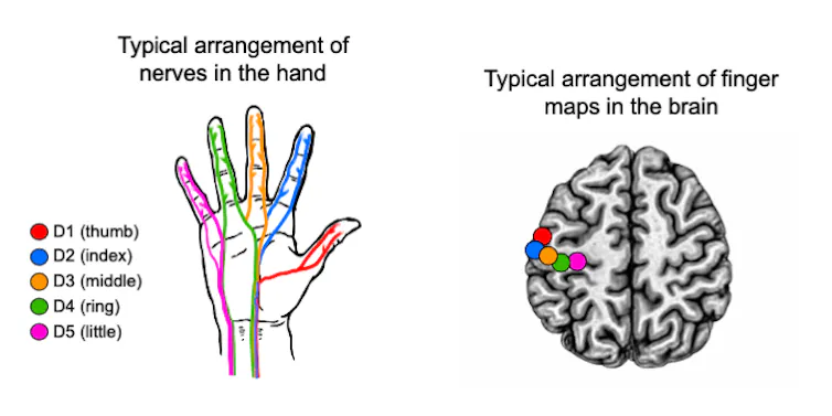
Ken Valyear/JohnBonachon/Wikimedia Commons (public domain), CC BY
When a repaired nerve grows back
Peripheral nerves – the network connecting your brain and spinal cord to the rest of your body – have an extraordinary capacity to regenerate. If one of the major nerves supplying the hand is cut, surgeons can reconnect its severed ends. New nerve fibres can grow back into the hand, allowing sensation and movement to return.
But nerve regeneration is not precisely guided. The growing fibres don’t receive instructions telling them exactly where to reconnect. They can therefore take different paths and connect with different parts of the hand from those they supplied before the injury.
Communication between the hand and brain can be restored, but the information reaching the brain may no longer have exactly the same organisation. Someone might touch their index finger but feel the sensation in the middle finger.
We know from experiments involving monkeys that nerve repair alters the brain’s hand map, changing the usual arrangement of responses to different fingers. These findings became influential in thinking about rehabilitation in humans, with some theories proposing that recovery might involve restoring a more normal hand map.
We used functional MRI to map brain responses to each finger being touched in 21 people who had undergone surgical repair of one or more major hand nerves, and compared them with 30 people without nerve injuries.
In people with repaired nerves, the normal organisation of responses to the different fingers was altered and more variable from person to person. The pattern resembled the irregular maps previously observed in monkeys after nerve repair.
The altered maps were not simply the result of weaker responses to touch. Quite the opposite: touching the repaired hand produced unusually strong responses in the relevant brain area.
The map changes could partly reflect how the regenerating nerves have reconnected in the hand. We didn’t directly measure those connections, however, and the unusually strong brain responses suggest that changes within the brain itself are also involved.
What does an altered map mean?
We still do not fully understand how the precise organisation of somatotopic maps contributes to perception and behaviour. Nerve repair gives a rare opportunity to study what happens when the normal relationship between the body and the brain changes.
We found that the map can change, but we did not find a clear relationship between the extent of that change and how well people could use their hands. People with more altered maps were not reliably worse at localising touch, nor did they show greater impairment on broader measures of hand function. That finding challenges a simple assumption: that a more “normal” brain map must necessarily be a more functional one.
Successful recovery may not require the brain to recreate its original map. Instead, the nervous system may learn to make effective use of a new relationship between the hand and brain.
Read more:
New MRI method could help track Huntington’s disease and its treatments
That could also be important for rehabilitation. Recovery after serious nerve injuries is often incomplete, with people experiencing lasting problems with sensation, movement or pain. Understanding how the brain adapts as a nerve regrows, and why recovery varies, will require following people from surgery through nerve regrowth and rehabilitation, tracking brain maps alongside behaviour and how the peripheral nerves reconnect.
That could ultimately tell us two things: how changes in the brain relate to recovery after nerve injury, and something more fundamental about the brain itself. For all the history and importance of the body’s maps in neuroscience, we still do not know exactly what their detailed organisation is for. By seeing what happens when that organisation changes, we may have a powerful way to investigate that question.

Ken Valyear was supported by a grant from the Wellcome Trust (215186/Z/19/Z).
Original source: https://analysis1.mil-osi.com/2026/09/14/new-insights-into-how-the-brain-responds-after-a-severed-nerve-is-repaired-could-boost-recovery/